

The kidney
How the kidneys filter blood, reclaim most of what they filter and secrete drugs into the urine; how kidney function is measured and staged; which hormones the kidney makes; and which supplement effects on the kidney have been documented in people.
ICRP Publication 89 (2002), Tables 2.39 and 2.40 and paragraphs 329, 380 and 383.
Status. Education, not medical advice. Figures marked as calculations are educational calculations from published equations, not measurements and not predictions for any person, product or ingredient. Reference ranges for laboratory tests vary by laboratory.
Related: ADME, PK and PBPK · Contaminants · Lessons learned
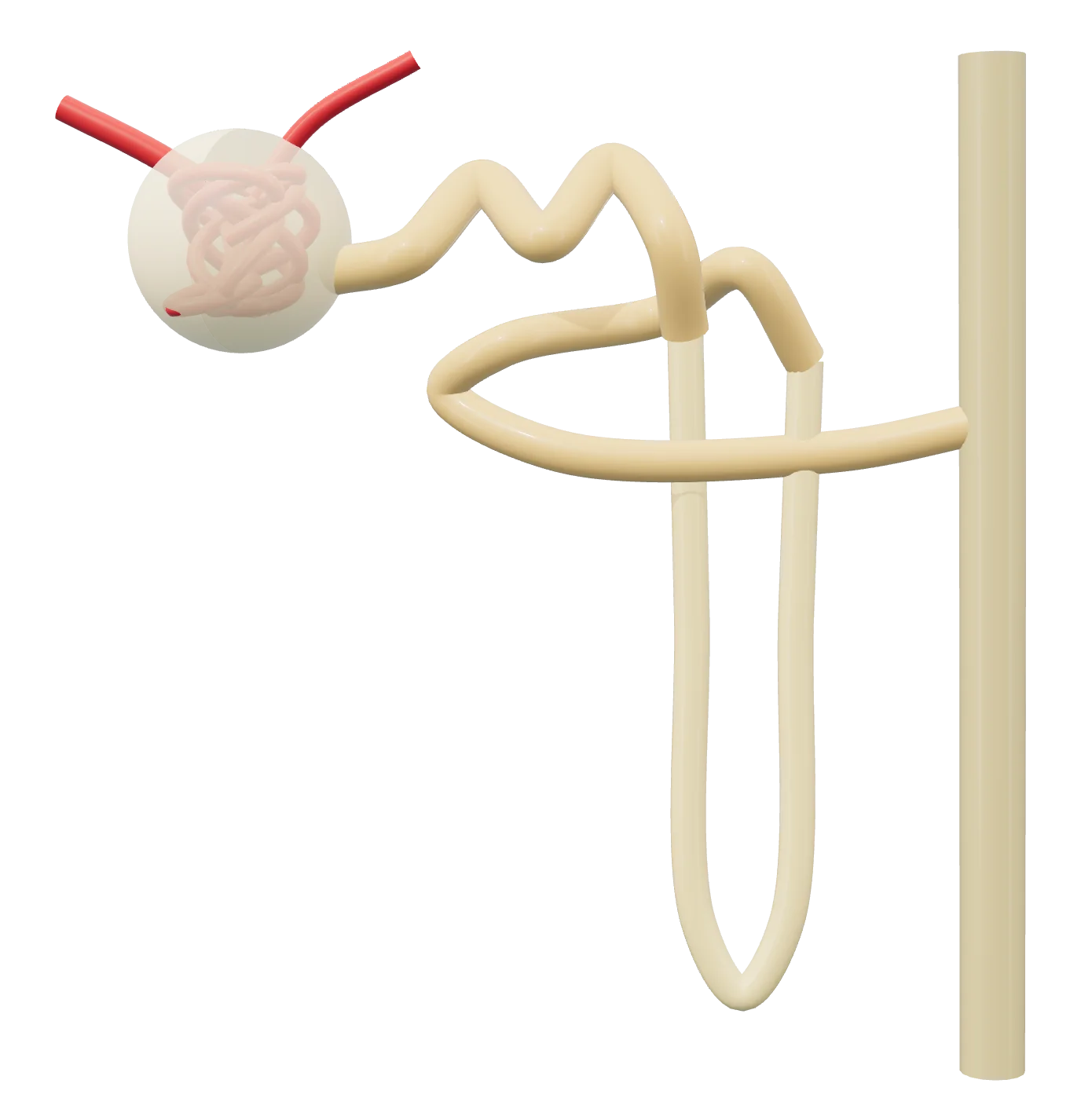
- 1 Glomerulus in Bowman’s capsule: blood is filtered
- 2 Proximal tubule: most water and solutes reabsorbed
- 3 Loop of Henle
- 4 Distal tubule
- 5 Collecting duct
Rendered by EUTECNIQ; generalized and not to scale.
Each nephron filters plasma, then its tubule takes most of the filtrate back
Each kidney has an outer cortex and an inner medulla and holds about one million nephrons. A nephron starts at the renal corpuscle, where a tuft of glomerular capillaries sits inside Bowman's capsule. Its tubule then runs through the proximal tubule, the loop of Henle and the distal convoluted tubule before draining into a collecting duct.
Blood reaches the glomerulus through the afferent arteriole and leaves through the efferent arteriole, which feeds a second capillary bed around the tubules. Those peritubular capillaries take up what the tubules reabsorb. At the glomerulus's vascular pole, macula densa cells in the tubule wall sense the salt content of the tubular fluid; with the two arterioles and the extraglomerular mesangium they form the juxtaglomerular apparatus.
Text version of the drawing: the seven numbered segments
- Renal corpuscleGlomerular capillaries inside Bowman's capsule, fed by the afferent arteriole and drained by the efferent arteriole. Filtration is about 125 mL/min (about 180 L/day) in a middle-aged man; solutes under about 5,000 Da are freely filtered unless bound to plasma proteins.
- Proximal tubuleConvoluted and straight parts. Reabsorbs about 65% of the filtrate, 65–70% of filtered sodium and about 90% of filtered glucose (through SGLT2). Secretes organic anions, organic cations and creatinine.
- Thin limbs of the loop of HenleThe descending limb penetrates the medulla and is highly permeable to water but not to solutes, so luminal osmolality rises toward the tip of the loop.
- Thick ascending limbReabsorbs 20–25% of filtered sodium through NKCC2 and is impermeable to water, so it dilutes the tubular fluid. The loop as a whole reabsorbs about 15% of the filtrate.
- Macula densaCells at the glomerular vascular pole, part of the juxtaglomerular apparatus. A fall in NaCl concentration in the tubular fluid signals renin release.
- Distal convoluted tubuleReabsorbs 5–7% of filtered sodium through the Na–Cl cotransporter NCC.
- Connecting tubule and collecting ductReabsorb 2–5% of filtered sodium through ENaC under aldosterone and secrete potassium. Aquaporin-2 water channels, moved to the cell membrane under vasopressin, let water follow the medullary osmotic gradient; urine can reach about 1,400 mosmol/L.
| Segment | Reabsorbs | Secretes | Main transport proteins |
|---|---|---|---|
| Proximal tubule | ≈65% of the filtrate; 65–70% of filtered Na+; ≈90% of filtered glucose; ≈65% of filtered K+ | Organic anions and cations, including many drugs; creatinine | NHE3 (Na+/H+ exchange), SGLT2; OAT1, OAT3, OCT2, MATE1, MATE2-K, P-gp, BCRP |
| Thin descending limb | Water | — | Water-permeable epithelium |
| Thick ascending limb | 20–25% of filtered Na+; ≈25% of filtered K+ (ICRP's “diluting segment”) | — | NKCC2; ROMK K+ channel |
| Distal convoluted tubule | 5–7% of filtered Na+ | — | NCC |
| Connecting tubule, collecting duct | 2–5% of filtered Na+; water when vasopressin is present | K+, increased by aldosterone | ENaC; aquaporin-2 |
Filtrate and potassium: ICRP Publication 89, paragraphs 383 and 389–391 (the potassium figure of 25% is given for the "diluting segment"). Sodium: Gembillo et al. 2026. NHE3: Zheng and Layton, Front Physiol 2026;17:1828410. Glucose, NKCC2, ROMK, NCC, ENaC: Gembillo et al. 2026. Thin limbs: Zacchia et al. 2018. Drug transporters: section 4. Published shares are rounded, come from different sources and need not add to 100%.
About a fifth of cardiac output passes through 310 grams of kidney
Renal blood flow is about 4 mL per gram of tissue per minute, far above most organs. About 18% of the plasma entering the kidneys is filtered; this ratio of GFR to renal plasma flow is the filtration fraction. Kidney transport work uses about 8% of the body's total oxygen consumption.
| Quantity | Value | ICRP 89 location |
|---|---|---|
| Mass of both kidneys | 310 g (adult male); 275 g (adult female) | Table 2.8 |
| Renal blood flow | 19% of cardiac output, male; 17%, female. With cardiac output 6.5 and 5.9 L/min: 1.24 and 1.00 L/min (computed) | Tables 2.39, 2.40 |
| Perfusion | About 4 mL blood per g tissue per min | Paragraph 378 |
| Glomerular filtration rate | About 125 mL/min, about 180 L/day (middle-aged adult male); lower on average in females; declines with age | Paragraph 380 |
| Renal plasma flow; filtration fraction | About 700 mL/min (young adult male); FF = GFR/RPF ≈ 0.18 | Paragraph 380 |
| Freely filtered solutes | Molecular mass under 5,000 Da, except those bound to plasma proteins; about half of plasma calcium is protein-bound | Paragraph 381 |
| Urine volume | Typically 1,200–2,000 mL/day; reference 1,600 mL/day (male), 1,200 mL/day (female) | Paragraph 385, Table 2.41 |
| Maximal urine concentration | About 1,400 mosmol/L; obligatory urine about 0.5 L/day | Paragraph 125 |
| Kidney blood content | 2.0% of total blood volume | Table 2.14 |
ICRP, 2002. Basic Anatomical and Physiological Data for Use in Radiological Protection: Reference Values. ICRP Publication 89. Organ blood flows for physiologically based models are tabulated on ADME, PK and PBPK.
Show data: share of the filtered load reabsorbed per segment
| Segment | Filtrate (water), % | Filtered Na+, % |
|---|---|---|
| Proximal tubule | 65 | 65–70 |
| Loop of Henle (Na+: thick ascending limb) | 15 | 20–25 |
| Distal tubule (Na+: distal convoluted tubule) | 10 | 5–7 |
| Collecting duct | 9.3 | 2–5 |
| Excreted in urine | 0.7 | about 1 |
| Substance | Filtered per minute | Excreted in urine per minute |
|---|---|---|
| Sodium | 18 mEq | 0.14 mEq |
| Potassium | 0.63 mEq | 0.066 mEq |
| Glucose | 125 mg | 0 mg |
| Urea | 31 mg | 18 mg |
| Uric acid | 3.8 mg | 0.42 mg |
| Creatinine | 1.4 mg | 1.2 mg |
ICRP Publication 89, Table 8.3 (modified from Guyton, 1982), assuming GFR 125 mL/min and urine flow 1.1 mL/min. ICRP gives about 60% of filtered urea and about 15% of filtered urate as excreted (paragraphs 400–401). These are textbook reference values, not measurements in one person.
Renal clearance adds filtration and secretion and subtracts reabsorption
Renal clearance is the volume of plasma the kidneys free of a substance per unit time. Only the unbound fraction of a drug in plasma is filtered, so filtration clearance is fu × GFR. A renal clearance clearly above that value means the tubules secrete the compound; a value below it means net reabsorption. The general pharmacokinetic equations are on ADME, PK and PBPK.
| Quantity | Equation or criterion | Source |
|---|---|---|
| Renal (urinary) clearance | CLR = (U × V̇) / P: urine concentration times urine flow rate, divided by plasma concentration | Provenzano et al., J Nephrol 2024 |
| Filtration fraction | FF = GFR / RPF | ICRP 89, paragraph 380 |
| Filtration clearance of a drug | CLGF = fu × GFR | Cristea et al., AAPS J 2020 |
| Active secretion clearance (no reabsorption assumed) | CLR − fu,p × GFR | ICH M12, section 2.2.1 |
| Model with filtration and active secretion | CLR = fu·GFR + (QR − GFR)·fu·CLint,sec / (QR + fu·CLint,sec/B:P) | Cristea et al. 2020 (no passive reabsorption or renal metabolism) |
| Secretion indicated | CLR > 1.5 × fu × GFR (assume fu = 0.01 if binding exceeds 99.9% or is unknown) | International Transporter Consortium, Giacomini et al. 2010 |
| Test as renal transporter substrate | Active renal secretion ≥ 25% of systemic clearance: study OAT1, OAT3, OCT2, MATE1 and MATE2-K in vitro | ICH M12, section 2.2.1 |
| Dedicated renal impairment study | Fraction of systemically available drug or active metabolite excreted unchanged in urine ≥ 0.3 | FDA guidance, March 2024 |
| eGFR in mL/min for dosing | eGFR (mL/min) = eGFR (mL/min/1.73 m²) × BSA / 1.73 | FDA guidance, March 2024 |
Provenzano M, et al. J Nephrol 2024;37:573–586; Cristea S, et al. AAPS J 2020;22:87; ICH M12 Drug Interaction Studies, adopted May 21, 2024 (issued by FDA as final guidance, docket FDA-2022-D-1527); Giacomini KM, et al. Nat Rev Drug Discov 2010;9:215–236; FDA, Pharmacokinetics in Patients with Impaired Renal Function — Study Design, Data Analysis, and Impact on Dosing, guidance for industry, March 2024.
Show data: calculated renal clearance, L/h
| CLint,sec, L/h | fu = 1 | fu = 0.5 | fu = 0.1 |
|---|---|---|---|
| 0.1 | 7.6 | 3.8 | 0.8 |
| 1 | 8.4 | 4.2 | 0.8 |
| 10 | 15.4 | 8.0 | 1.6 |
| 100 | 45.8 | 30.6 | 8.7 |
| 1,000 | 69.5 | 61.8 | 39.0 |
| 10,000 | 73.6 | 69.4 | 62.8 |
Uptake transporters on the blood side and efflux transporters on the urine side move drugs through the proximal tubule
Active secretion is a two-step relay through the proximal tubule cell. OAT1, OAT3 and OCT2 take compounds up from blood across the basolateral membrane; MATE1, MATE2-K and the ATP-driven pumps P-gp and BCRP push them into the urine across the apical brush border. URAT1 works the other way, reabsorbing urate from the urine.
Regulators ask drug developers to test new drugs against these transporters. ICH M12 asks for inhibition studies on P-gp, BCRP, OATP1B1, OATP1B3, OAT1, OAT3, OCT2, MATE1 and MATE2-K. OATP4C1 and URAT1 are not on that list; they appear in the International Transporter Consortium's map of the proximal tubule.
| Transporter | Gene | Membrane and direction | Example substrates | Example inhibitors |
|---|---|---|---|---|
| OAT1 | SLC22A6 | Basolateral; blood into cell | Adefovir, p-aminohippurate, cidofovir, tenofovir (in vitro) | Probenecid, benzylpenicillin (in vitro, OAT1/OAT3) |
| OAT3 | SLC22A8 | Basolateral; blood into cell | Benzylpenicillin, estrone-3-sulfate, methotrexate, pravastatin (in vitro); clinical: furosemide, methotrexate | Probenecid (clinical, OAT1/OAT3) |
| OCT2 | SLC22A2 | Basolateral; blood into cell | Creatinine, metformin, tetraethylammonium (in vitro) | Cimetidine, clonidine (in vitro); clinical: cimetidine, dolutegravir |
| OATP4C1 | SLCO4C1 | Basolateral in the ITC map; blood into cell (UniProt instead describes an apical uptake role, inferred by similarity) | Digoxin, ouabain, methotrexate, thyroid hormones T3 and T4 (UniProt) | Not listed by FDA |
| MATE1 | SLC47A1 | Apical (brush border); cell into urine, in exchange for H+ | Creatinine, metformin, MPP+, tetraethylammonium (in vitro) | Cimetidine, pyrimethamine (in vitro); clinical: trimethoprim, pyrimethamine |
| MATE2-K | SLC47A2 | Apical (brush border); cell into urine, in exchange for H+ | As MATE1 (in vitro) | As MATE1 |
| P-gp | ABCB1 | Apical; cell into urine, ATP-driven | Digoxin, fexofenadine, loperamide, quinidine (in vitro) | Cyclosporine, ketoconazole, quinidine, verapamil (in vitro) |
| BCRP | ABCG2 | Apical (brush border); cell into urine, ATP-driven | Estrone-3-sulfate, methotrexate, rosuvastatin, sulfasalazine (in vitro) | Elacridar, Ko143, novobiocin (in vitro) |
| URAT1 | SLC22A12 | Apical; urine into cell (reabsorption) | Urate, exchanged for lactate, nicotinate or chloride (UniProt) | Not listed by FDA |
FDA, Drug Development and Drug Interactions: Table of Substrates, Inhibitors and Inducers, Tables 3-1 and 3-2 (content current June 5, 2023); FDA, Healthcare professionals: examples of drugs that interact with CYP enzymes and transporter systems, Table 1 (content current May 29, 2026); UniProt; Giacomini et al. 2010; Huls et al. 2008. The ITC notes that the membrane localization of some transporters is controversial. "In vitro" marks FDA's examples for laboratory studies; "clinical" marks drugs FDA lists from human interaction studies.
| Transporter | ICH M12 cutoff to exclude clinical inhibition risk |
|---|---|
| OAT1, OAT3, OCT2 | IC50,u > 10 × Cmax,u (Cmax,u/IC50,u < 0.1) |
| MATE1, MATE2-K | IC50,u > 50 × Cmax,u (Cmax,u/IC50,u < 0.02) |
| P-gp, BCRP (oral drugs) | IC50,u > 0.1 × (Dose/250 mL) |
ICH M12, Table 1. Cmax,u: unbound maximal plasma concentration of the inhibitor at steady state after the therapeutic dose. M12 also lists endogenous biomarkers: N1-methylnicotinamide and N1-methyladenosine for OCT2, MATE1 and MATE2-K, and pyridoxic acid for OAT1/3.
Creatinine is itself an OCT2 and MATE substrate, and drugs that inhibit its secretion can raise serum creatinine. KDIGO lists decreased tubular secretion among the non-GFR determinants of serum creatinine, which is why such a rise does not by itself show a fall in filtration.
Show data: decreases in renal clearance
| Interacting drug → affected drug | Likely transporter | Reported effect |
|---|---|---|
| Probenecid → furosemide | OATs | CLR −66% |
| Probenecid → cidofovir | OATs | CLR −32% |
| Probenecid → acyclovir | OATs | CLR −32%, AUC +40% |
| Cimetidine → metformin | OCTs | CLR −27%, AUC +50% |
| Cimetidine → dofetilide | OCTs | CLR −33% |
| Cimetidine → pilsicainide | OCTs | CLR −28% (AUC +33%) in one study, −41% in another |
| Quinidine → digoxin | P-gp | CLR −34% to −48% |
Serum creatinine becomes an estimate of GFR only after age and sex enter the equation
Measuring GFR needs the plasma or urinary clearance of an exogenous marker such as iohexol, iothalamate or 51Cr-EDTA, usually with repeated blood samples, and is available only in certain centers. Laboratories instead report an estimated GFR (eGFR) calculated from a blood marker made by the body. KDIGO recommends a validated estimating equation over the serum marker alone, and advises against using race in the calculation.
| Test | What it is | Affected by more than GFR |
|---|---|---|
| Serum creatinine | Made in muscle from creatine phosphate; filtered, not reabsorbed, and secreted in the late proximal nephron, so excretion runs about 10% above filtration. Adult reference excretion 1.7 g/day (male), 1.0 g/day (female) | Muscle mass and diet (meat, high-protein diets, creatine supplements), tubular secretion and drugs that block it, extrarenal elimination. Men had 21% higher serum creatinine than women in a 9-year study of 144 subjects |
| Cystatin C | A 120-amino-acid cysteine protease inhibitor found in plasma and other body fluids (UniProt P01034) | Thought to be higher adiposity, smoking, hypo- and hyperthyroidism, glucocorticoid excess and chronic inflammation; less understood than for creatinine |
| Blood urea nitrogen (BUN) | Urea nitrogen; the liver converts ammonia from protein breakdown into urea, which the kidneys excrete. About 60% of filtered urea reaches the urine | Higher with dehydration, burns, some medicines, a high-protein diet or a recent heart attack; lower with low protein intake, malnutrition or liver disease. Levels vary with sex and usually rise with age |
| Urine albumin-to-creatinine ratio (ACR) | Albumin in a urine sample divided by urine creatinine, in mg/g or mg/mmol; a marker of kidney damage | Not a GFR test. KDIGO categories: A1 <30, A2 30–300 (moderately increased relative to the young adult level), A3 >300 mg/g |
Creatinine and urea: ICRP Publication 89, paragraphs 397–400 and Table 2.42. Measured GFR and non-GFR determinants: KDIGO 2024 CKD guideline, Tables 7, 8 and 9 and chapter 1.2 text. Cystatin C protein: UniProt P01034 (146-residue precursor with a 26-residue signal peptide). BUN: MedlinePlus, BUN (Blood Urea Nitrogen) test, updated October 27, 2024. ACR: KDIGO 2024, Table 3. Numerical reference ranges vary by laboratory and are not given here.
The CKD-EPI 2021 creatinine equation, as published
eGFRcr = 142 × min(Scr/κ, 1)α × max(Scr/κ, 1)−1.200 × 0.9938Age × 1.012 [if female]
κ = 0.7 (female) or 0.9 (male); α = −0.241 (female) or −0.302 (male); Scr = serum creatinine in mg/dL (multiply µmol/L by 1/88.4); Age in years; eGFR in mL/min per 1.73 m² of body-surface area. There is no race coefficient.
Inker LA, Eneanya ND, Coresh J, et al., for the Chronic Kidney Disease Epidemiology Collaboration. New creatinine- and cystatin C–based equations to estimate GFR without race. N Engl J Med 2021;385:1737–1749, Table 2 ("2021 CKD-EPI creatinine": intercept 142; a1 F −0.241, M −0.302; a2 −1.200; age coefficient 0.9938; female 1.012) and its footnote formula. Conversion factor 88.4 µmol/L per mg/dL: the same paper and KDIGO 2024.
eGFRcr-cys = 135 × min(Scr/κ, 1)α × max(Scr/κ, 1)−0.544 × min(Scys/0.8, 1)−0.323 × max(Scys/0.8, 1)−0.778 × 0.9961Age × 0.963 [if female]
κ as above; α = −0.219 (female) or −0.144 (male); Scys = serum cystatin C in mg/L.
Inker et al. 2021, Table 2 ("2021 CKD-EPI creatinine–cystatin C"). In their validation set of 4,050 people, 85% or more of eGFR values were within 30% of measured GFR for every equation tested, and the new creatinine–cystatin C equations were more accurate than the new creatinine equations. KDIGO 2024 recommends eGFRcr-cys where eGFRcr is less accurate and GFR affects a clinical decision (Recommendation 1.2.2.1, grade 1C).
Show data: calculated eGFR, mL/min/1.73 m²
| Serum creatinine, mg/dL | Woman 30 | Man 30 | Woman 50 | Man 50 | Woman 70 | Man 70 |
|---|---|---|---|---|---|---|
| 0.6 | 124 | 133 | 109 | 118 | 97 | 104 |
| 0.8 | 102 | 122 | 90 | 108 | 79 | 95 |
| 1.0 | 78 | 104 | 69 | 92 | 61 | 81 |
| 1.2 | 62 | 83 | 55 | 74 | 49 | 65 |
| 1.5 | 48 | 64 | 42 | 56 | 37 | 50 |
| 2.0 | 34 | 45 | 30 | 40 | 26 | 35 |
| 3.0 | 21 | 28 | 18 | 25 | 16 | 22 |
| 4.0 | 15 | 20 | 13 | 17 | 11 | 15 |
For drug dosing, FDA asks that eGFR be converted from mL/min per 1.73 m² to mL/min with the person's body-surface area, because a drug's renal clearance follows the individual's own GFR.
KDIGO stages chronic kidney disease by GFR and albuminuria together
KDIGO defines chronic kidney disease (CKD) as abnormalities of kidney structure or function present for at least 3 months, with implications for health. Either a GFR below 60 mL/min/1.73 m² or a marker of kidney damage, such as an ACR of 30 mg/g or more, qualifies. CKD is classified by cause, GFR category (G1–G5) and albuminuria category (A1–A3).
Show data: the KDIGO categories in text
| GFR category | GFR, mL/min/1.73 m² | Term | A1 (<30 mg/g) | A2 (30–300 mg/g) | A3 (>300 mg/g) |
|---|---|---|---|---|---|
| G1 | ≥90 | Normal or high | Low | Moderately increased | High |
| G2 | 60–89 | Mildly decreased | Low | Moderately increased | High |
| G3a | 45–59 | Mildly to moderately decreased | Moderately increased | High | Very high |
| G3b | 30–44 | Moderately to severely decreased | High | Very high | Very high |
| G4 | 15–29 | Severely decreased | Very high | Very high | Very high |
| G5 | <15 | Kidney failure | Very high | Very high | Very high |
Without evidence of kidney damage, neither G1 nor G2 meets the criteria for CKD. G2 "mildly decreased" and A2 "moderately increased" are relative to the young adult level.
FDA uses a separate classification only to enroll dedicated renal impairment studies of drugs: normal ≥90, mild 60 to <90, moderate 30 to <60, and severe impairment or kidney failure not on dialysis <30 mL/min. FDA states it is not meant for classifying kidney disease.
KDIGO 2024, Tables 1–3; FDA, Pharmacokinetics in Patients with Impaired Renal Function, March 2024, Table 1.
The kidney also makes renin, erythropoietin and calcitriol
Renin starts a cascade that raises blood pressure and sodium retention. Juxtaglomerular cells at the tips of the afferent arterioles release it when perfusion pressure falls, when circulating or sympathetic-nerve catecholamines stimulate their β-adrenergic receptors, or when the macula densa detects less NaCl. Angiotensin II then acts directly on vascular smooth muscle as a vasoconstrictor and stimulates aldosterone release from the adrenal cortex.
Text version of the cascade
Angiotensinogen (from the liver) is cut by renin to angiotensin I, which angiotensin-converting enzyme (ACE) converts to angiotensin II. Angiotensin II acts on angiotensin receptors (AGTR1 and AGTR2) as a direct vasoconstrictor and stimulates the zona glomerulosa of the adrenal cortex to make aldosterone. Aldosterone increases sodium reabsorption through ENaC and potassium secretion in the connecting tubule and collecting duct.
| Hormone | Made by | Main stimulus | What it does |
|---|---|---|---|
| Renin (enzyme, EC 3.4.23.15) | Juxtaglomerular cells at the tips of the afferent arterioles | Fall in perfusion pressure; β-adrenergic stimulation; low NaCl at the macula densa | Cuts angiotensinogen to angiotensin I, its only known function, starting the cascade above |
| Erythropoietin (EPO) | Peritubular interstitial fibroblasts of the kidney, the primary source in adults; the liver is the main site during embryonic development | Hypoxia, through hypoxia-inducible factors (HIF) | Binds its receptor EPOR to drive red blood cell production; inadequate EPO production contributes to the anemia of chronic kidney disease |
| Calcitriol (1α,25-dihydroxyvitamin D3) | The kidney, by 25-hydroxyvitamin D-1α-hydroxylase (CYP27B1, EC 1.14.15.18) | Regulation not covered on this page | The hormonally active form of vitamin D; acts through the vitamin D receptor in calcium and phosphorus homeostasis |
Yamaguchi et al. 2023; UniProt P00797, P01588 (EPO), O15528 (CYP27B1), retrieved October 29, 2026; Haase VH. Regulation of erythropoiesis by hypoxia-inducible factors. Blood Rev 2013;27:41–53; ICRP Publication 89, paragraph 334 (the kidney's two endocrine functions, calcitriol and erythropoietin).
Five supplement links to the kidney documented in people
Each item below rests on human data or on an agency statement. Some change a kidney test without a change in measured GFR; others are documented harms. None of this is advice about whether to take a supplement.
Creatine can raise serum creatinine without lowering measured GFR
KDIGO's table of indications for cystatin C lists high-protein diets and creatine supplements as non-GFR determinants of serum creatinine, noting minimal data. In a 20-year-old man with a single kidney who took creatine (20 g/day for 5 days, then 5 g/day for 30 days) on a high-protein diet, serum creatinine rose while 51Cr-EDTA clearance did not change. A 12-week randomized, placebo-controlled trial in people with type 2 diabetes found no significant difference in 51Cr-EDTA clearance between creatine and placebo.
| Measure | Before | After 35 days |
|---|---|---|
| Serum creatinine, mg/dL | 1.03 | 1.27 |
| Measured GFR, 51Cr-EDTA clearance, mL/min/1.73 m² | 81.6 | 82.0 |
| eGFR from serum creatinine, CKD-EPI 2021 (calculated on this page) | 106.6 | 82.9 |
KDIGO 2024, Table 8; Gualano B, et al. Am J Kidney Dis 2010;55:e7–e9 (case report); Gualano B, et al. Eur J Appl Physiol 2011;111:749–756 (randomized trial; 51Cr-EDTA clearance difference −0.3 mL/min/1.73 m², 95% CI −24.9 to 24.2); Williamson L, New D. BMJ Case Rep 2014 (creatine ethyl ester raised serum creatinine in the absence of kidney pathology; it reversed after stopping). The eGFR row is an educational calculation from the published inputs (male, age 20), not a value reported by the authors. Related: Creatine.
Vitamin C intake was linked to more kidney stones in men
In three U.S. cohorts followed for a median 11.3 to 11.7 years, 6,245 incident kidney stones occurred. Higher total and supplemental vitamin C intake was associated with higher stone risk in men, not in women; dietary vitamin C was not associated in either. The authors lacked stone composition for all cases. NIH's Office of Dietary Supplements notes that high intakes can raise urinary oxalate and that the best evidence of a role in stone formation is in people with pre-existing hyperoxaluria.
Show data: hazard ratios in men
| Vitamin C intake | Hazard ratio (95% CI) |
|---|---|
| Total <90 mg/day | 1.00 (reference) |
| Total 90–249 mg/day | 1.19 (0.99–1.46) |
| Total 250–499 mg/day | 1.15 (0.93–1.42) |
| Total 500–999 mg/day (median ≈700) | 1.29 (1.04–1.60) |
| Total ≥1,000 mg/day | 1.43 (1.15–1.79) |
| Supplemental ≥1,000 mg/day vs no use | 1.19 (1.01–1.40) |
Ferraro et al. 2016; NIH Office of Dietary Supplements, Vitamin C fact sheet for health professionals, updated July 31, 2025 (adult UL 2,000 mg/day). Related: Vitamin C.
Aristolochic acid can cause kidney failure and urothelial cancer
In Belgium, slimming pills dispensed by one clinic in 1990–1992 contained Aristolochia fangchi in place of Stephania tetrandra. FDA's May 31, 2000 letter reported over 100 patients with nephropathy and at least 70 who needed a kidney transplant or dialysis. Among 39 patients with end-stage disease who had their native kidneys and ureters removed, 18 had urothelial carcinoma (46%; 95% CI 29–62%).
| Body | Statement | Date |
|---|---|---|
| FDA, letter to health care professionals | Concern about botanical products, including dietary supplements, containing aristolochic acid; names Aristolochia, Asarum, Bragantia, Stephania, Clematis, Akebia, Cocculus, Diploclisia, Menispermum and Sinomenium, and trade names such as Mu tong and Fang ji | May 31, 2000 (attachments updated April 2001) |
| FDA Import Alert 54-10 | Detention without physical examination of dietary supplements containing aristolochic acid; "a potent carcinogen and nephrotoxin" | Published December 10, 2020 |
| IARC Monographs Volume 100A | Aristolochic acid and plants containing aristolochic acid: carcinogenic to humans (Group 1) | 2012 |
Nortier JL, et al. N Engl J Med 2000;342:1686–1692; FDA, Letter to Health Care Professionals: FDA Concerned About Botanical Products, Including Dietary Supplements, Containing Aristolochic Acid, May 31, 2000 (archived copy); FDA Import Alert 54-10; IARC Monographs Volume 100A, Plants Containing Aristolochic Acid, section 5. More: Contaminants · Lessons learned.
Potassium and magnesium accumulate when the kidneys cannot excrete them
NIH's Office of Dietary Supplements states that the potassium intake recommendations do not apply to people whose potassium excretion is impaired by kidney disease or by medicines. Potassium-sparing diuretics, ACE inhibitors and angiotensin receptor blockers reduce urinary potassium excretion and can cause hyperkalemia, and experts recommend monitoring potassium in people taking them, especially with impaired kidney function. In people with normal kidney function, ODS reports no evidence that high potassium intakes cause hyperkalemia.
The kidneys largely control magnesium balance and excrete about 120 mg per day in urine. ODS states that the risk of magnesium toxicity rises with impaired renal function or kidney failure, because the ability to remove excess magnesium is reduced or lost. Very large doses of magnesium-containing laxatives and antacids, typically above 5,000 mg/day, have been associated with magnesium toxicity. The adult upper level for supplemental magnesium is 350 mg/day.
NIH Office of Dietary Supplements, Potassium fact sheet for health professionals, updated June 2, 2022; Magnesium fact sheet for health professionals, updated January 6, 2026. Related: Potassium · Magnesium.
Licorice lowers plasma potassium by blocking the inactivation of cortisol
Glycyrrhetic acid, from the glycyrrhizinic acid in licorice root, inhibits 11β-hydroxysteroid dehydrogenase type 2, the enzyme that inactivates cortisol to cortisone. Cortisol then occupies mineralocorticoid receptors, causing sodium and water retention and potassium loss, with renin and aldosterone suppressed. This pseudohyperaldosteronism can show as edema, hypokalemia and raised blood pressure.
A meta-analysis of 18 studies (337 participants) taking at least 100 mg of glycyrrhizic acid daily found a mean fall in plasma potassium of 0.33 mmol/L, a rise in systolic blood pressure of 5.45 mm Hg (95% CI 3.51–7.39), and falls in plasma renin activity and aldosterone. The European Scientific Committee on Food set 100 mg/day as an upper limit for regular ingestion and noted it may not protect people with hypertension or reduced enzyme activity. NCCIH states that even small amounts of glycyrrhizin from licorice root products have been linked to severe adverse effects in people with hypertension or with heart or kidney conditions.
European Commission, Scientific Committee on Food. Opinion on glycyrrhizinic acid and its ammonium salt, SCF/CS/ADD/EDUL/225 Final, April 10, 2003; Penninkilampi R, Eslick EM, Eslick GD. J Hum Hypertens 2017;31:699–707; NCCIH, Licorice Root, last updated April 2025. Related: Licorice root.
Where these topics continue
Related pages: ADME, PK and PBPK (pharmacokinetic equations, PBPK structure, organ blood flows) · Contaminants · Lessons learned · Creatine · Vitamin C · Potassium · Magnesium · Licorice root · Sodium and electrolytes
Health information notice
These statements have not been evaluated by the Food and Drug Administration. Nothing on this site is intended to diagnose, treat, cure or prevent any disease. The information is for general education only. It is not medical advice and does not replace advice, diagnosis or treatment from a qualified health professional, or any medication a clinician has prescribed.
Do not start, stop or change any medicine, supplement or herbal product because of something you read here. Talk to your doctor or pharmacist first, especially if you are pregnant or breastfeeding, have a medical condition, take any medicines, are preparing for surgery, or are caring for a child. Research findings, traditional uses and safety data are reported as their sources state them; they are not recommendations or claims about any product. If you think you may have a medical emergency, call your doctor or 911 at once.
EUTECNIQ Sciences sells no supplements or health products today and receives nothing from any product or ingredient mentioned on this site; any commercial interest it later has in an ingredient will be stated on that ingredient’s page.