

Microorganisms in botanicals
Plant materials carry bacteria, yeasts and molds from soil, water, animals and people, and a few of them cause infection or form toxins. This page lists those organisms, the pharmacopeial limits that apply to them, and how laboratories count, detect and identify them.
USP 35–NF 30 ⟨2023⟩ Table 2; FDA BAM Chapter 3 (January 2026), revision history; Stevenson et al., ISME J 2015; Hindson et al., Anal Chem 2011.
Status. EUTECNIQ is in formation. It operates no microbiology laboratory and has tested no samples; methods here are described from their published texts.
Course: Microbiology of botanicals covers kill steps, acid tolerance, biofilms, cells that do not form colonies, and has plate-count and growth-window calculators. Related: mycotoxins and other contaminants · process hygiene · quality system
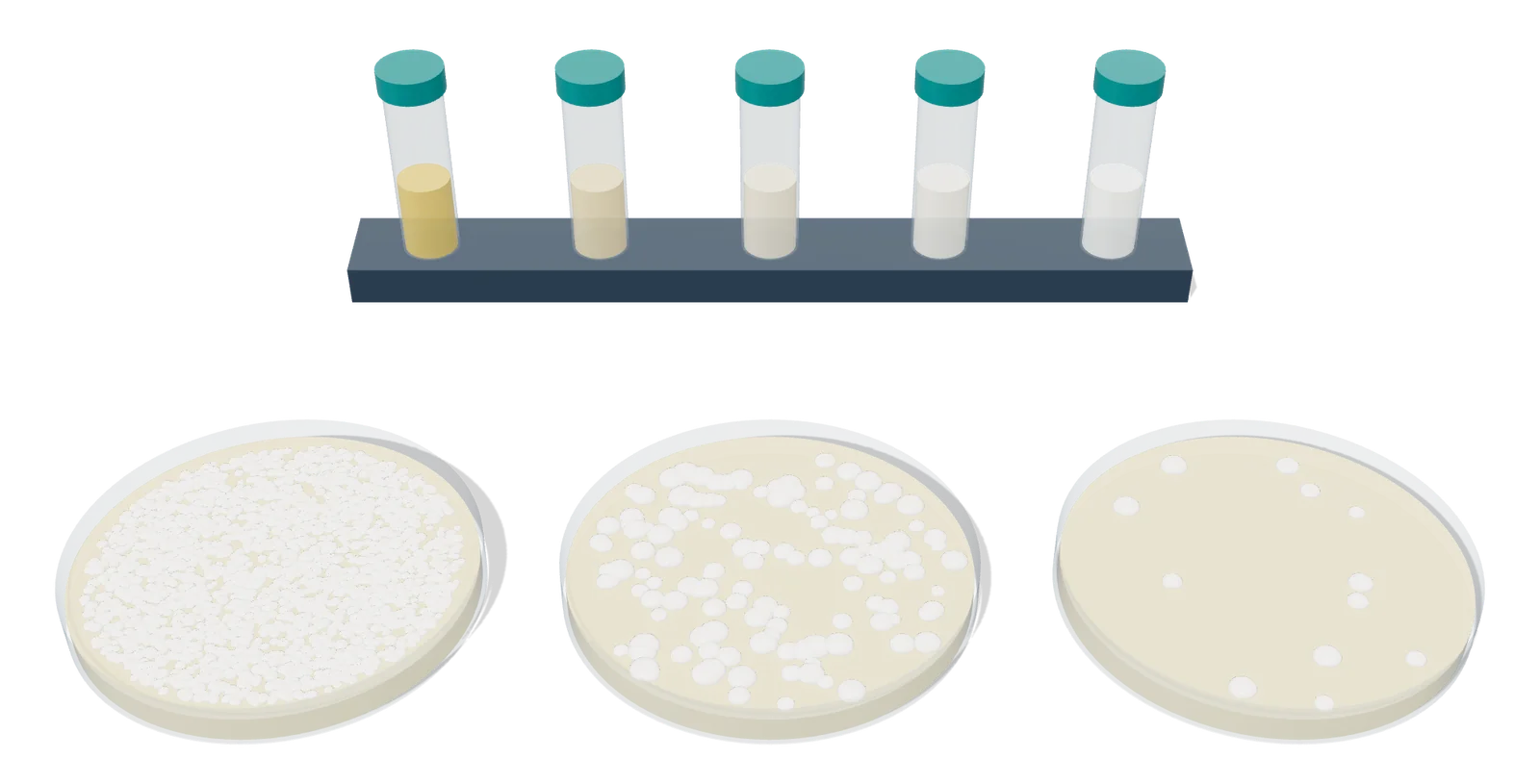
- 1 Tenfold serial dilution
- 2 Spread plates after incubation
Tenfold serial dilution and spread plates, rendered by EUTECNIQ; colony numbers are illustrative.
Bacteria of concern
FDA's Bad Bug Book records Bacillus cereus in soil and vegetation, Listeria monocytogenes in soil and decaying vegetation, and Cronobacter in herbs and spices; Salmonella survives well in spices. Some of these organisms cause infection. Others harm through a toxin: Staphylococcus aureus enterotoxins and the emetic toxin of B. cereus form in the food, and C. botulinum neurotoxin forms during growth.
Two groups in the table are not single pathogens. E. coli and the bile-tolerant Gram-negative bacteria mark contamination and hygiene; Pseudomonas aeruginosa and the Burkholderia cepacia complex matter mainly in water-based liquids and topical products.
| Organism | Gram stain and form | Where it occurs | Hazard | Limit or test |
|---|---|---|---|---|
| Salmonella spp. | Gram-negative rod; no spores; Enterobacteriaceae | Survives in low-moisture foods; spices have carried large outbreaks | Infection | Absent in 10 g (USP ⟨2023⟩); absent in 25 g (Ph. Eur. 5.1.8); FDA BAM Ch. 5 |
| Escherichia coli | Gram-negative rod; Enterobacteriaceae | Intestine of humans and warm-blooded animals; proposed as an indicator of fecal contamination in 1892 | Indicator; most strains not pathogens | Absent in 10 g (USP ⟨2023⟩); absent in 1 g, or 10³ cfu/g for herbal teas (Ph. Eur.) |
| Shiga toxin-producing E. coli | Gram-negative rod | Outbreaks traced to lettuce, spinach, sprouts and unpasteurized juice | Infection; Shiga toxins | No USP ⟨2023⟩ criterion; FDA BAM Ch. 4A |
| Staphylococcus aureus | Gram-positive cocci in grape-like clusters | Nose, throat, hair and skin of 50% or more of healthy people; food handlers | Heat-stable enterotoxins formed in food | Tested in USP ⟨2022⟩; no ⟨2023⟩ criterion; not detected in 10 g (NSF/ANSI 173) |
| Bacillus cereus | Gram-positive large rod; facultatively anaerobic; endospores | Soil and vegetation | Diarrheal toxin made in the intestine; emetic toxin (cereulide) made in food | No USP ⟨2023⟩ criterion; FDA BAM Ch. 14 |
| Clostridium perfringens | Gram-positive rod; anaerobic but aerotolerant; heat-resistant spores | Soil, sediments and animal intestines | Enterotoxin | Clostridia test with an 80 °C, 10 min heated portion (USP ⟨2022⟩, ⟨62⟩); FDA BAM Ch. 16 |
| Clostridium botulinum | Gram-positive rod; anaerobic; heat-resistant spores | Cultivated and forest soils; sediments | Neurotoxin formed during growth | No count limit; growth controlled by water activity, pH, temperature and oxygen (next sections) |
| Listeria monocytogenes | Gram-positive rod; facultative; motile | Soil, decaying vegetation, moist environments; persists in food plants; grows below 1 °C | Infection; among the leading causes of death from foodborne illness | No USP ⟨2023⟩ criterion; FDA BAM Ch. 10 |
| Cronobacter spp. | Gram-negative motile rod | Isolated from herbs, spices, cereals and rice; survives up to 2 years in powdered infant formula (aw about 0.2) | Infection, chiefly in infants | FDA BAM Ch. 29 |
| Bile-tolerant Gram-negative bacteria | Gram-negative; includes Enterobacteriaceae | Group test, not one species | Hygiene indicator | 10³ cfu/g, dried botanicals (USP ⟨2023⟩); 10² or 10⁴ (Ph. Eur. B or C); USP ⟨62⟩ |
| Pseudomonas aeruginosa | Gram-negative; forms biofilms | Soil and water; most human- or animal-impacted environments | Opportunistic infection | Absent in 1 g or 1 mL for cutaneous, oromucosal and nasal drug products (USP ⟨1111⟩); cetrimide agar (⟨62⟩) |
| Burkholderia cepacia complex | Gram-negative group of species | Survives or multiplies in non-sterile water-based products; resists some preservatives | Opportunistic infection, especially in immunocompromised people | USP ⟨60⟩ (published December 1, 2019), which FDA recommends |
FDA Bad Bug Book, 2nd ed. (2012): Salmonella, EHEC, S. aureus, B. cereus, C. perfringens, C. botulinum, L. monocytogenes and Cronobacter chapters; FDA BAM Ch. 4 (October 2020), introduction; USP 35–NF 30 ⟨2022⟩, ⟨2023⟩, ⟨62⟩, ⟨1111⟩; Ph. Eur. 5.1.8 (10th ed.); AHPA comparison of microbial limits (June 2022) for NSF/ANSI 173; Diggle and Whiteley, Microbiology 2020; FDA statement on B. cepacia complex in non-sterile water-based drugs.
Yeasts and molds
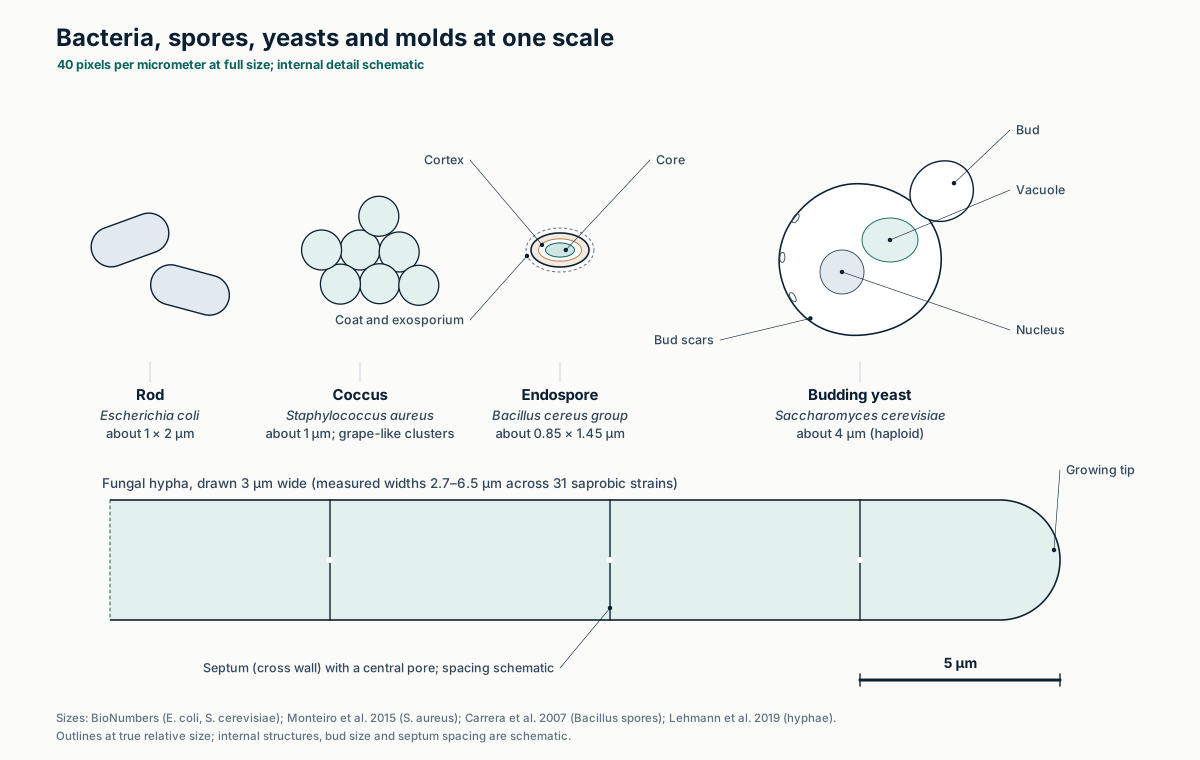
FDA's Bacteriological Analytical Manual notes that most foodborne molds can grow at a water activity of 0.85 or less, while yeasts generally need more water, and that most mycotoxins are stable compounds not destroyed by processing. A yeast-and-mold count therefore says nothing about toxin already formed; mycotoxins need their own assays, covered on the contaminants page.
| Genus | Mycotoxins named for the genus | Counting note |
|---|---|---|
| Aspergillus | Aflatoxins (A. flavus, A. parasiticus); ochratoxin A; patulin | Dichloran 18% glycerol agar (aw 0.955) preferred when the sample's aw is below 0.95 |
| Penicillium | Ochratoxin A; patulin | Experienced analysts identify it on the plate at 10–30× magnification; malt extract agar for identification (FDA BAM Ch. 18) |
| Fusarium | Deoxynivalenol, nivalenol, T-2 and HT-2 toxins, zearalenone, fumonisins | Common in soil (WHO) |
| Alternaria | Alternariol, alternariol monomethyl ether, tenuazonic acid, altertoxins, tentoxin | EFSA found the toxins in grains, tomato products, sunflower seeds and fruit |
| Mucor | None among the toxins WHO lists | A "spreader" that can overgrow plates; dichloran and rose bengal in DRBC agar slow it |
| Yeasts | None in WHO's list; FDA BAM says "possibly yeasts" | Counted with molds as TYMC; generally need higher water activity than molds |
FDA BAM Ch. 18, Yeasts, Molds and Mycotoxins (April 2001), introduction and media appendix; WHO fact sheet, Mycotoxins (October 2, 2023); EFSA CONTAM Panel, Alternaria toxins in feed and food, EFSA J 2011;9(10):2407.
Spores and vegetative cells
Bacillus and Clostridium species can form endospores, dormant cells that survive drying and heat that kill growing (vegetative) cells. The pharmacopeial Clostridia test uses this: one portion of the sample is heated to 80 °C for 10 min and cooled, the other is not, and both are cultured anaerobically.
| Feature of the endospore | Effect |
|---|---|
| Core water content much lower than in a growing cell | Main determinant of wet-heat resistance |
| α/β-type small acid-soluble proteins saturate the DNA | Protect DNA against wet heat, dry heat and UV |
| Large pool of dipicolinic acid in the core | Contributes to UV resistance |
| Killed by dry heat, UV and gamma radiation through DNA damage | Kill steps for spores need far more than for vegetative cells; see the Academy course |
Setlow P, J Appl Microbiol 2006;101:514–525 (Bacillus subtilis spores); USP 35–NF 30 ⟨2022⟩ and ⟨62⟩, Clostridia.
Water activity and growth
Each pathogen has a minimum water activity (aw) below which it does not grow. FDA's table measured each limit with salt as the humectant and the other factors near optimal, so combined stresses narrow the windows. No growth is not death: Salmonella and Cronobacter survive in dry foods, and USP ⟨2023⟩ says only that low-aw products "will not be susceptible to microbial growth during their shelf life."
| Organism | Min. aw | Min. pH | Growth range, °C | Oxygen | Spores |
|---|---|---|---|---|---|
| C. botulinum type E, non-proteolytic B and F | 0.97 | 5.0 | 3.3–45 | Anaerobe | Yes |
| Pathogenic E. coli | 0.95 | 4.0 | 6.5–49.4 | Facultative | No |
| Salmonella spp. | 0.94 | 3.7 | 5.2–46.2 | Facultative | No |
| C. botulinum type A, proteolytic B and F | 0.935 | 4.6 | 10–48 | Anaerobe | Yes |
| C. perfringens | 0.93 | 5.0 | 10–52 | Anaerobe | Yes |
| B. cereus | 0.92 | 4.3 | 4–55 | Facultative | Yes |
| L. monocytogenes | 0.92 | 4.4 | −0.4–45 | Facultative | No |
| S. aureus, toxin formation | 0.85 | 4.0 | 10–48 | Facultative | No |
| S. aureus, growth | 0.83 | 4.0 | 7–50 | Facultative | No |
| Most foodborne molds | ≤ 0.85 | 2 | 10–35, a few beyond | Mostly aerobic | Fungal spores |
| Xerophilic A. penicillioides, X. bisporus | 0.640 | – | – | – | Fungal spores |
FDA, Fish and Fishery Products Hazards and Controls Guidance, Appendix 4, Table A-1 (bacteria; aw with salt); FDA BAM Ch. 18 (molds: aw 0.85 or less, pH 2 to above 9, 10–35 °C); Stevenson et al., ISME J 2015;9:1333–1351 (0.640, with mixed solutes; earlier limit about 0.65); spore formers as named in the FDA Bad Bug Book; the others listed do not form spores. The Academy course has an interactive growth-window explorer built on Table A-1.
Pharmacopeial microbial limits
USP ⟨2023⟩ recommends limits by how processed a material is, and applies them only when the monograph is silent. It states that coliform or Enterobacteriaceae counts "will not be an appropriate general microbiological criterion for botanicals." Ph. Eur. 5.1.8 uses three categories; its maximum acceptable count is five times the stated criterion.
| Material | TAMC, NMT | TYMC, NMT | BTGN, NMT | Absence required |
|---|---|---|---|---|
| Dried or powdered botanicals | 10⁵ | 10³ | 10³ | Salmonella and E. coli in 10 g |
| Powdered botanical extracts | 10⁴ | 10³ | – | Salmonella and E. coli in 10 g |
| Tinctures | 10⁴ | 10³ | – | – |
| Fluidextracts | 10⁴ | 10³ | – | – |
| Infusions and decoctions | 10² | 10 | – | – |
| Nutritional supplements with botanicals | 10⁴ | 10³ | – | Salmonella and E. coli in 10 g |
| Botanicals to be treated with boiling water before use | 10⁵ | 10³ | – | E. coli in 10 g |
| Other raw materials and supplement ingredients (Table 3) | 10³ | 10² | Not specified | E. coli in 10 g |
| Supplements with synthetic or highly refined ingredients (Table 3) | 10³ | 10² | Not specified | E. coli in 10 g |
USP 35–NF 30, ⟨2023⟩ Tables 2 and 3; same values for dried botanicals, extracts and supplements in AHPA's June 2022 comparison. Later editions revised the boiling-water row (compared on the Academy page); USP lists 2025 versions of ⟨2021⟩–⟨2023⟩, so confirm the current official text in USP–NF. TAMC: total aerobic microbial count; TYMC: total combined yeasts and molds count; BTGN: bile-tolerant Gram-negative bacteria; NMT: not more than.
| Category | TAMC | TYMC | BTGN | E. coli | Salmonella | S. aureus |
|---|---|---|---|---|---|---|
| Ph. Eur. A: herbal teas used with boiling water | 10⁷ (max 5 × 10⁷) | 10⁵ (max 5 × 10⁵) | – | 10³ cfu/g | Absent in 25 g | – |
| Ph. Eur. B: extracts and pre-treated herbal drugs | 10⁴ | 10² | 10² | Absent in 1 g | Absent in 25 g | – |
| Ph. Eur. C: where processing cannot meet category B | 10⁵ | 10⁴ | 10⁴ | Absent in 1 g | Absent in 25 g | – |
| NSF/ANSI 173, finished product, non-extract | 10⁷ | 10⁵ | 10⁴ | 10² cfu/g* | Not detected in 10 g | Not detected in 10 g |
| NSF/ANSI 173, finished product, extract | 10⁴ | 10³ | 10² | Not detected in 10 g | Not detected in 10 g | Not detected in 10 g |
Ph. Eur. 10th ed., 5.1.8 (category descriptions abbreviated; maximum = 5 × criterion). NSF/ANSI 173 values as listed in AHPA, Recommended Microbial Limits for Botanical Ingredients, comparison table (June 2022); *if E. coli is confirmed, colonies are tested for enterovirulent E. coli, which has zero tolerance. For drug products, harmonized USP ⟨61⟩ reads a criterion of 10¹ cfu as a maximum acceptable count of 20, 10² as 200, and so on.
What the USP tests do
The supplement chapters ⟨2021⟩ and ⟨2022⟩ count and detect; the drug chapters ⟨61⟩ and ⟨62⟩ do the same with harmonized media and incubation. Before any count is trusted, a suitability test spikes the sample with known organisms and checks they are recovered, because many botanicals inhibit growth.
| Chapter | Purpose | Media and incubation | Readout and suitability |
|---|---|---|---|
| ⟨2021⟩ (USP 35–NF 30) | TAMC and TYMC of supplements; 10 g or 10 mL brought to 100 mL (1:10) | Soybean–casein digest agar, 30–35 °C, 48–72 h; Sabouraud dextrose agar, 20–25 °C, 5–7 days | Dilute to 30–300 colonies per 1 mL; blank 1:10 plates reported as fewer than 10 per g; more than 70% recovery in preparatory testing |
| ⟨61⟩ (harmonized, official May 1, 2025) | TAMC and TYMC of nonsterile drug products | Membrane filtration (pore ≤ 0.45 µm), pour plate (1 mL in 15–20 mL agar) or spread plate (≥ 0.1 mL); 30–35 °C, 3–5 days and 20–25 °C, 5–7 days | Plates under 250 (TAMC) or 50 (TYMC) colonies; inoculum ≤ 100 cfu; recovery within a factor of 2; MPN only for TAMC when no other method works |
| ⟨2022⟩ (USP 35–NF 30) | Absence of Salmonella, E. coli, S. aureus and Clostridium; 10 g, with an optional 25 g retest of a doubtful result | Salmonella: Rappaport–Vassiliadis broth, then brilliant green, XLD or Hektoen agar. E. coli: MacConkey broth at 42–44 °C, then MacConkey agar. Clostridium: 80 °C for 10 min, reinforced clostridial medium, anaerobic, 35–37 °C, 48 h | Suspect colonies confirmed by identification tests |
| ⟨62⟩ (harmonized) | Bile-tolerant Gram-negatives, E. coli, Salmonella, P. aeruginosa, S. aureus, Clostridia, Candida albicans | Enterobacteria enrichment broth Mossel then violet red bile glucose agar; cetrimide agar; mannitol salt agar; Columbia agar (anaerobic); Sabouraud media; mostly 30–35 °C | S. aureus: yellow or white colonies with a yellow zone; Salmonella: red colonies on XLD, with or without black centers |
USP 35–NF 30 ⟨2021⟩, ⟨2022⟩, ⟨62⟩; USP ⟨61⟩ Stage 4 harmonized text, official May 1, 2025 (USP harmonization page).
Plate counts
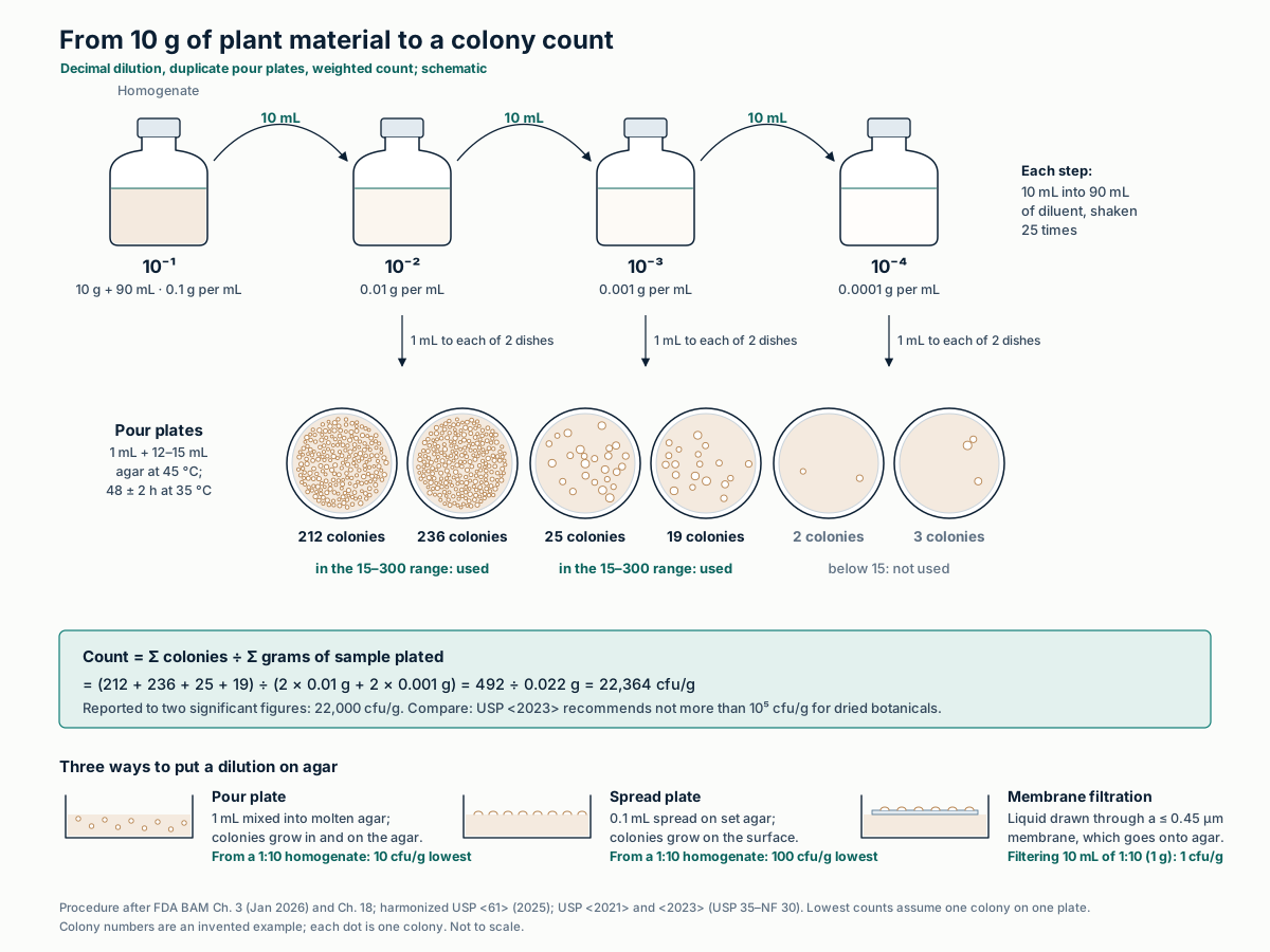
A colony-forming unit (cfu) is any cell or clump that grows into one visible colony. The sample is diluted in tenfold steps until a plate holds a countable number. FDA's estimate is a weighted average: total colonies on all countable plates divided by the total amount of sample those plates received. Results are rounded to two significant figures.
| Method | Countable colonies per plate |
|---|---|
| FDA BAM aerobic plate count (pour plate, since March 2025) | 15–300 |
| ISO 4833, as cited by FDA BAM | 15–300 |
| APHA, as cited by FDA BAM; BAM before March 2025 | 25–250 |
| USDA and AOAC, as cited by FDA BAM | 30–300 |
| USP ⟨2021⟩ (USP 35–NF 30) | 30–300 |
| Harmonized USP ⟨61⟩: TAMC / TYMC | < 250 / < 50 |
| FDA BAM yeasts and molds (spread plate, 25 °C, 5 days) | 10–150 |
| Spiral plater, one inoculation (per mL of sample) | 500–500,000 |
FDA BAM Ch. 3, Aerobic Plate Count (January 2026), introduction, Sections C–D and the spiral plate method; FDA BAM Ch. 18; USP 35–NF 30 ⟨2021⟩; harmonized USP ⟨61⟩ (2025).
Most probable number and detection
The most probable number (MPN) method inoculates replicate tubes at several dilutions and records which grow. It assumes random distribution and that any tube receiving one viable cell turns positive; the MPN is the concentration λ that makes the observed pattern most likely, solving Σ gjmj/(1 − e−λmj) = Σ tjmj. FDA notes it is particularly useful below about 100 organisms per g.
The same Poisson model sets what a presence or absence test can see: the chance that a portion of mass m holds at least one cell is 1 − e−cm. A negative 10 g test is consistent with a lot at 0.1 cfu/g about 37% of the time.
| Positive tubes | MPN/g, solved here | MPN/g, BAM Table 1 | BAM 95% interval |
|---|---|---|---|
| 1-0-0 | 3.57 | 3.6 | 0.17–18 |
| 2-1-0 | 14.7 | 15 | 3.7–42 |
| 3-0-0 | 23.1 | 23 | 4.6–94 |
| 3-1-0 | 42.7 | 43 | 9–180 |
| 3-2-0 | 93.3 | 93 | 18–420 |
FDA BAM Appendix 2, Most Probable Number from Serial Dilutions (August 2023), introduction and Table 1. Solved by bisection on the likelihood equation; BAM rounds to two significant figures.
Enrichment and selective media
Detection methods for pathogens run in stages: a nonselective pre-enrichment, selective broths that favor the target over competitors, differential agars on which its colonies look distinct, then screening slants and identification tests. FDA dilutes allspice, cinnamon and oregano 1:100 and cloves 1:1000, because no method neutralizes their toxicity to Salmonella culture.
| Medium | Target | Reaction described by the method |
|---|---|---|
| Xylose lysine desoxycholate (XLD) agar | Salmonella | Pink colonies with or without black centers (BAM); red with or without black centers (USP ⟨62⟩) |
| Hektoen enteric agar | Salmonella | Blue-green to blue colonies with or without black centers |
| Bismuth sulfite agar | Salmonella | Brown, gray or black colonies, sometimes with a metallic sheen |
| Triple sugar iron and lysine iron agar slants | Salmonella screening | Alkaline (red) slant and acid (yellow) butt in TSI, with or without H₂S; alkaline (purple) butt in LIA |
| MacConkey broth at 42–44 °C | E. coli | Growth subcultured to MacConkey agar; colonies need identification |
| Violet red bile glucose agar | Bile-tolerant Gram-negatives | Any growth counts |
| Cetrimide agar | P. aeruginosa | Growth indicates possible presence; confirm |
| Mannitol salt agar | S. aureus | Yellow or white colonies surrounded by a yellow zone |
| DRBC and DG18 agars | Molds and yeasts | DRBC slows spreading molds; DG18 (aw 0.955) for samples below aw 0.95 |
FDA BAM Ch. 5 (October 2026), Section E.7–E.9; FDA BAM Ch. 18 media appendix; USP 35–NF 30 ⟨2022⟩ and ⟨62⟩.
Real-time and digital PCR
Real-time PCR (qPCR) reads fluorescence every cycle. The threshold cycle (Ct) falls by about 3.3 cycles per tenfold more target at 100% efficiency; a standard curve gives the efficiency, E = 10−1/slope − 1. PCR also detects DNA of dead cells unless a dye such as propidium monoazide removes it. FDA uses qPCR or LAMP to screen the 24 h Salmonella pre-enrichment: negatives are reported negative, positives go to culture. Validated qPCR matrices include black and white pepper, cumin, paprika, basil and oregano.
Droplet digital PCR splits a 20 µL reaction into about 20,000 droplets and counts positive droplets at the end. With p the positive fraction, copies per droplet λ = −ln(1 − p), with no standard curve.
| Positive droplets | p | λ = −ln(1 − p), copies per droplet | Copies per reaction (× 20,000) |
|---|---|---|---|
| 20 | 0.001 | 0.0010 | 20 |
| 4,000 | 0.20 | 0.223 | 4,463 |
| 12,000 | 0.60 | 0.916 | 18,326 |
| 19,980 | 0.999 | 6.91 | 138,155 |
Hindson et al., Anal Chem 2011;83:8604–8610 (about 20,000 droplets of about 1 nL from 20 µL; Poisson correction; range from one copy to about 100,000 copies). At p = 0.999 the estimate rests on 20 negative droplets and is imprecise. Nocker et al., J Microbiol Methods 2006 (propidium monoazide). FDA BAM Ch. 5, Section D and Appendix A.
Rapid screens and identification
ATP bioluminescence measures all ATP present, microbial or not, so on plant material it reports plant residue as well as microbes. MALDI-TOF identifies an isolated colony by matching its protein mass fingerprint to a database. Sequencing reads marker genes or whole genomes. Each needs the growth or enrichment step before it, which sets most of the time to result.
| Method | What it measures | Resolution and speed | Main limitation |
|---|---|---|---|
| ATP bioluminescence | Light from luciferase acting on ATP, in relative light units | Immediate; no identification | Cannot separate microbial from non-microbial ATP |
| MALDI-TOF mass spectrometry | Protein fingerprint over m/z of about 2,000–20,000 (proteins of about 2–20 kDa), mostly ribosomal proteins | Genus or species of an isolate in minutes | Only organisms whose spectra are in the database |
| 16S rRNA gene sequencing | About 1,500 bp gene; 500–525 bp minimum for diagnostic use | Genus in over 90% of isolates; species in 65–83%; species call needs more than 99% similarity | Poor resolution in some groups, including Enterobacteriaceae and B. cereus / B. anthracis |
| ITS sequencing (fungi) | Nuclear ribosomal internal transcribed spacer | Highest probability of correct identification across fungi of six regions tested | Weaker in some groups; supplementary barcodes proposed |
| Amplicon sequencing of a sample | 16S or ITS reads from all DNA in the sample | Community profile without culture | Host (plant) sequences compete; PCR clamps can block them. Counts DNA, not live cells |
| Whole-genome sequencing | Complete genome of an isolate | FDA uses it with SeqSero to assign Salmonella serotypes | Needs a pure isolate |
Kotsiou et al., Antimicrob Resist Infect Control 2026;15:44; Singhal et al., Front Microbiol 2015;6:791; Janda and Abbott, J Clin Microbiol 2007;45:2761–2764; Schoch et al., PNAS 2012;109:6241–6246; Lundberg et al., Nat Methods 2013;10:999–1002; FDA BAM Ch. 5, Section F.
Validating alternative methods
A rapid method replaces a compendial or reference method only after it is shown to give equivalent or better results in the matrix tested. USP ⟨1223⟩ asks whether the alternative "will yield results equivalent to, or better than" the compendial method and sets the parameters by test type. ISO 16140-2 gives the protocol for food-chain methods, and FDA requires verification for any matrix not already validated.
| Parameter | Qualitative (presence or absence) | Quantitative (counts) |
|---|---|---|
| Accuracy, precision, quantification limit, linearity, operational range | No | Yes |
| Specificity, detection limit, robustness, repeatability, ruggedness | Yes | Yes |
| Framework | Scope | Key requirement |
|---|---|---|
| USP ⟨1223⟩ | Alternatives to USP microbiological tests | Counts at least as high as the compendial method, or not less than 70% of its estimate; overlapping 95% intervals for detection limits |
| ISO 16140-2:2016, Amd 1:2024 | Alternative, mostly proprietary, methods for microbiology in the food chain | Method comparison study and interlaboratory study, for qualitative and quantitative methods; the amendment revises relative level of detection and relative trueness calculations |
| AOAC Performance Tested Methods | Proprietary test kits | Developer study plus an independent laboratory study, expert review, annual renewal |
| FDA Foods Program | FDA laboratory methods | Verification or validation under FDA's microbiology method validation guidelines for matrices not listed as validated |
USP 35–NF 30 ⟨1223⟩, Table 1 and text (the chapter has since been revised; check current USP–NF); ISO 16140-2:2016 scope (preview of EN ISO 16140-2) and ISO 16140-2:2016/Amd 1:2024 title; AOAC Research Institute, Performance Tested Methods program page; FDA BAM Ch. 5, Section D.
Where EUTECNIQ would work
Proposed EUTECNIQ's planned laboratory would run compendial counts and absence tests with documented method suitability for each botanical, and would adopt rapid methods only after verification in that matrix. Until then, testing would be placed with qualified laboratories.
Related: Academy: Microbiology of botanicals · Process hygiene · Contaminants and mycotoxins · Quality system · Reading a certificate of analysis
Health information notice
These statements have not been evaluated by the Food and Drug Administration. Nothing on this site is intended to diagnose, treat, cure or prevent any disease. The information is for general education only. It is not medical advice and does not replace advice, diagnosis or treatment from a qualified health professional, or any medication a clinician has prescribed.
Do not start, stop or change any medicine, supplement or herbal product because of something you read here. Talk to your doctor or pharmacist first, especially if you are pregnant or breastfeeding, have a medical condition, take any medicines, are preparing for surgery, or are caring for a child. Research findings, traditional uses and safety data are reported as their sources state them; they are not recommendations or claims about any product. If you think you may have a medical emergency, call your doctor or 911 at once.
EUTECNIQ Sciences sells no supplements or health products today and receives nothing from any product or ingredient mentioned on this site; any commercial interest it later has in an ingredient will be stated on that ingredient’s page.